HEK-Blue™ mNOD1 Cells
-
Cat.code:
hkb-mnod1
- Documents
HEK 293 cells express endogenous TLR3 and TLR5 levels.
ABOUT
Murine NOD1 expressing HEK293 reporter cells
HEK-Blue™ mNOD1 cells were engineered from the human embryonic kidney HEK 293 cell line to assess the role of the Nucleotide-binding Oligomerization Domain-containing protein 1 (NOD1).
These cells stably express the murine (m) NOD1 gene and an NF-κB-inducible SEAP reporter gene. SEAP (secreted embryonic alkaline phosphatase) activity upon NOD1 stimulation can be readily determined by performing the assay in HEK-Blue™ Detection. This cell culture medium allows for real-time detection of SEAP. Alternatively, SEAP activity may be monitored using QUANTI-Blue™, a SEAP detection reagent.
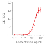
The overexpression of mNOD1 renders HEK-Blue™ mNOD1 cells to strongly respond to NOD1-specific ligands (see figures). As HEK293 cells express endogenous levels of TLR3 and TLR5 [in-house data], HEK-Blue™ mNOD1 cells will respond to their cognate ligands such as Poly(I:C), and flagellin. They do not express human NOD2 (see figure).
NOD1 and NOD2 are cytosolic pattern recognition receptors (PRRs), specialized to sense distinct motifs of peptidoglycan, an essential constituent of the bacterial cell wall. NOD1 senses the D-γ-glutamyl-meso-DAP dipeptide (iE-DAP), which is found in PGN of all Gram-negative and certain Gram-positive bacteria [1].
Key Features:
- Verified overexpression of murine NOD1
- Strong and stable response to NOD1-specific ligands
- Distinct monitoring of NF-κB by assessing the SEAP activities
Applications:
- Defining the distinct role of NOD1 in the NF-κB-dependent pathway
- Screening for NOD1-specific agonists or inhibitors in comparison with their parental cell line HEK-Blue™ Null1
Reference:
1. Correa RG, Milutinovic S, Reed JC,. 2012. Roles of NOD1 (NLRC1) and NOD2 (NLRC2) in innate immunity and inflammatory diseases. Biosci Rep. 32(6):597-608.
Disclaimer: These cells are for internal research use only and are covered by a Limited Use License (See Terms and Conditions). Additional rights may be available.
SPECIFICATIONS
Specifications
murine NOD1 activation cellular assays
Complete DMEM (see TDS)
Validated using PlasmotestTM.
Each lot is functionally tested and validated.
CONTENTS
Contents
-
Product:HEK-Blue™ mNOD1 Cells
-
Cat code:hkb-mnod1
-
Quantity:3-7 x 10^6 cells
- 1 ml of Blasticidin (10 mg/ml)
- 1 ml of Zeocin® (100mg/ml)
- 1 ml of Normocin™ (50 mg/ml)
- 1 pouch of HEK-Blue™ Detection
Shipping & Storage
- Shipping method: Dry ice
- Liquid nitrogen vapor
Storage:
Details
NOD1 and NOD2
The cytosolic NOD-Like Receptors (NLRs, also known as NODs or NALP) are Nucleotide-binding Oligomerization Domain containing receptors. To date, 22 NLRs have been identified in humans and constitute a major class of intracellular pattern recognition receptors (PRRs) [1].
The founding NLR-family members NOD1 (CARD4) and NOD2 (CARD15) recognize distinct motifs of peptidoglycan (PGN), an essential constituent of the bacterial cell wall. NOD1 senses the D-γ-glutamyl-meso-DAP dipeptide (iE-DAP), which is found in PGN of all Gram-negative and certain Gram-positive bacteria [1, 2] whereas NOD2 recognizes the muramyl dipeptide (MDP) structure found in almost all bacteria. Thus NOD2 acts as a general sensor of PGN and NOD1 is involved in the recognition of a specific subset of bacteria. Both iE-DAP and MDP must be delivered intracellularly either by bacteria that invade the cell or through other cellular uptake mechanisms. Ligand-bound NOD1 and NOD2 oligomerize and signal via the serine/threonine RIP2 kinase through CARD-CARD homophilic interactions [3]. Once activated, RIP2 mediates ubiquitination of NEMO/IKKγ leading to the activation of NF-κB and the production of inflammatory cytokines. Furthermore, poly-ubiquitinated RIP2 recruits TAK1, which leads to IKK complex activation and the activation of MAPKs [4].
Genetic mutations in NOD2 are associated with Crohn’s disease, a chronic inflammatory bowel disease [5]. In addition, numerous studies have recently revealed that NOD1 and NOD2 have a close relationship with a variety of cancers via controlling proliferation, altering immunosurveillance, and interacting with tissue bacteria, including intestinal commensal intestinal microflora. Moreover, additional research into the mechanisms of NOD1 and NOD2 in cancers would shed light on the innate immunity-cancer relationship and provide intriguing targets for immunotherapy [6].
References:
1. Chamaillard M. et al., 2003. An essential role for NOD1 in host recognition of bacterial peptidoglycan containing diaminopimelic acid. Nat. Immunol. 4: 702-707.
2. Girardin S. et al., 2003. Nod1 detects a unique muropeptide from Gram-negative bacterial peptidoglycan. Science 300: 1584-1587.
3. Kobayash, K. et al., 2002. RICK/Rip2/CARDIAK mediates signalling for receptors of the innate and adaptive immune systems. Nature 416: 194-199.
4. Kobayashi K. et al., 2005. Nod2-dependent regulation of innate and adaptive immunity in the intestinal tract. Science 307: 731-734.
5. Ogura Y. et al., 2001. A frameshift mutation in NOD2 associated with susceptibility to Crohn’s disease. Nature 411: 603-606.
6. Wang D, 2022. NOD1 and NOD2 Are Potential Therapeutic Targets for Cancer Immunotherapy. Comput Intell Neurosci.;2022:2271788.
DOCUMENTS
Documents
Technical Data Sheet
Safety Data Sheet
Validation Data Sheet
Certificate of analysis
Need a CoA ?


