The inflammasome is a large multiprotein complex which plays a key role in innate immunity by participating in the production of the pro-inflammatory cytokines interleukin-1β (IL-1β) and IL-18. These related cytokines cause a wide variety of biological effects associated with infection, inflammation and autoimmune processes.
They are both produced as inactive precursors, pro-IL-1β and pro-IL-18, and share a common maturation mechanism that requires caspase-1. Caspase-1 itself is synthesized as a zymogen, pro-caspase-1, that undergoes autocatalytic processing resulting in two subunits that form the active caspase-1. Activation of caspase-1 occurs within the inflammasome following its assembly.
NLRP3 inflammasome
The best characterized inflammasome is the NLRP3 (also known as NALP3 and cryopyrin) inflammasome. It comprises the NLR protein NLRP3, the adapter ASC and pro-caspase-1. The general consensus is that maturation and release of IL-1β requires two distinct signals: the first signal leads to synthesis of pro-IL-1β and other components of the inflammasome, such as NLRP3 itself; the second signal results in the assembly of the NLRP3 inflammasome, caspase-1 activation and IL-1β secretion.
Inflammasome Inducers
Activation of the NRLP3 inflammasome can be triggered by numerous stimuli, chemically and structurally highly different.
Microbial molecules (pathogen associated molecular patterns, PAMPs), such as bacterial lipopolysaccharide and fungal zymosan, can activate the NLRP3 inflammasome and induce IL-1β secretion in the presence of ATP [1]. External ATP, considered as a danger signal, causes the opening of the P2X7 receptor and the subsequent recruitment of the channel pannexin-1 leading to the release of intracellular potassium. The bacterial toxin nigericin has also been reported to induce the activation of NLRP3 by causing potassium efflux in a pannexin-1-dependent manner [2].
Besides PAMPs, the NLRP3 inflammasome can be activated by molecules associated with stress or danger, including crystalline and particulate substances. Crystals of uric acid and calcium pyrophosphatedihydrate, the aetiological agents of gout and pseudogout respectively, were the first crystals shown to engage the NLRP3 inflammasome [3]. Then, asbestos and silica were demonstrated to cause inflammatory lung diseases through a similar mechanism [4]. This mechanism is also responsible for the adjuvant properties of alum [5] and the pathogenicity of fibrillar amyloid-β, a particulate substance associated with Alzheimer’s disease [6]. Recently, malarial hemozoin, a heme crystal, was shown to act as a danger signal that activates the NLRP3 inflammasome [7].
Assembly of the NLRP3 inflammasome
Considering the differences in the structure and function of the molecules reported to activate the NLRP3 inflammasome, it is unlikely that they directly interact with NLRP3. Several mechanisms seem to play a role in the assembly of the NLRP3 inflammasome. Membrane damage appears to be a common step in NLRP3 activation shared by a number of stimuli including ATP, nigericin and crystals.
ATP and nigericin cause membrane damage by inducing the formation of a pore [2], while crystals create lysosomal membrane damage following their phagocytosis [5]. One hypothesis to explain how membrane damage can trigger NLRP3 activation is that this damage causes the modification or release of an endogenous molecule that is recognized by NLRP3. Another event that appears to be required for the activation of NLRP3 is the efflux of intracellular potassium.
Indeed, glyburide (also known as glybenclamide), an inhibitor of ATP-sensitive K+ channels, was shown to block the activation of the NLRP3 inflammasome in reponse to ATP, nigericin and crystals [8]. Lastly, the generation of reactive oxygen species (ROS) seems also critical for the activation of the NLRP3 inflammasome [7].
Inflammasone and the immune response
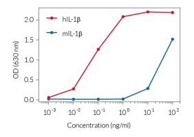
The increasing number of publications on the inflammasome highlight its importance in the immune response to microbial molecules and danger signals. The inflammasome is becoming an attractive target for therapeutic intervention in a wide range of inflammatory diseases, including autoimmune diseases. However, more studies need to be carried out to better understand the activation and regulation of the inflammasome. In an effort to assist scientists in their studies on the inflammasome, InvivoGen provides a set of tools comprising a cell line specifically enginereed to detect mature IL-1β, and molecules that act as inducers or inhibitors of the inflammasome.
References
1. Lamkanfi M. et al., 2009. Fungal zymosan and mannan activate the cryopyrin inflammasome. J Biol Chem. 284(31):20574-81.
2. Pelegrin P, Surprenant A., 2007. Pannexin-1 couples to maitotoxin- and nigericin-induced interleukin-1beta release through a dye uptake-independent pathway. J Biol Chem. 282(4):2386-94.
3. Martinon F. et al., 2006. Gout-associated uric acid crystals activate the NALP3 inflammasome. Nature. 440(7081):237-41.
4. Dostert C. et al., 2008. Innate immune activation through Nalp3 inflammasome sensing of asbestos and silica. Science. 320(5876):674-7.
5. Hornung v, et al., 2008. Silica crystals and aluminum salts activate the NALP3 inflammasome through phagosomal destabilization. Nat Immunol. 9(8):847-56.
6. Halle A. et al., 2008. The NALP3 inflammasome is involved in the innate immune response to amyloid-beta. Nat Immunol. 9(8):857-65.
7. Dostert C. et al., 2009. Malarial hemozoin is a Nalp3 inflammasome activating danger signal. PLoS One. 4(8):e6510.
8. Lamkanfi M. et al., 2009. Glyburide inhibits the Cryopyrin/Nalp3 inflammasome. J Cell Biol. 187(1):61-70.


