Anti-mTIGIT-mIgG2a InvivoFit™
-
Cat.code:
mtigit-mab10-1
- Documents
ABOUT
Recombinant murinized TIGIT antibody for in vivo use
Anti-mTIGIT-mIgG2a InvivoFit™ is a recombinant murinized anti-mouse (m)TIGIT monoclonal antibody (mAb) featuring the variable region of the previously described 10A7 clone [1] and a mouse (m)IgG2a constant region.
TIGIT (T cell immunoglobulin and ITIM domain) is a cell surface inhibitory immune checkpoint [1]. It is specifically expressed on immune cells including natural killer (NK) cells, activated and memory T cells, as well as regulatory T cells (Tregs) [2].
The 10A7 mAb has been used to block TIGIT signaling in Tregs and chronically stimulated CD8+ T cells [3, 4]. Using recombinant technology, the original 10A7 hamster IgG2a constant region has been replaced with a murine IgG2a format which mediates potent cytotoxic functions [5]. This murinization also allows for reduced immunogenicity of the administered mAb and risks of fatal hypersensitivity reactions [6-8].
Anti-mTIGIT-mIgG2a is provided in an InvivoFit™ grade, a high-quality standard specifically adapted to in vivo studies.
Key features of Anti-mTIGIT-mIgG2a InvivoFit™:
- Derives from the 10A7 clone, Hamster IgG2a
- Features the mIgG2a isotype (constant region)
- Filter-sterilized (0.2 µm), endotoxin level < 1 EU/mg
- Suitable for parental delivery in mice (azide-free)
- Low aggregation < 5%
- Produced in animal-free facilities and defined media
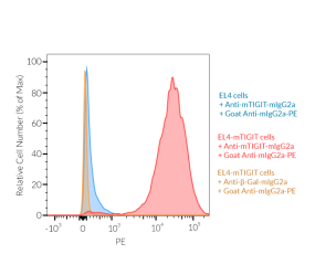
Anti-mTIGIT-mIgG2a InvivoFit™ is produced in Chinese hamster ovary (CHO) cells, purified by affinity chromatography with protein A, and its binding to mTIGIT is validated using flow cytometry.
References:
1. Yu X. et al, 2009. The surface protein TIGIT suppresses T cell activation by promoting the generation of mature immunoregulatory dendritic cells. Nat Immunol. 10(1):48-57.
2. Chauvin J.M. & Zarour H.M., 2020. TIGIT in cancer immunotherapy. J. Immunother. Cancer 8:e000957.
3. Jonhston R.J., et al, 2014. The immunoreceptor TIGIT regulates antitumor and antiviral CD8(+) T cell effector function. Cancer Cell. 26(6):923-937.
4. Joller, N. et al, 2014. Treg Cells Expressing the Coinhibitory Molecule TIGIT Selectively Inhibit Proinflammatory Th1 and Th17 Cell Responses. Immunity. 40(4):569-581.
5. Nimmerjahn F. & Ravetch J.V., 2005. Divergent immunoglobulin g subclass activity through selective Fc receptor binding. Science. 310:1510-2.
6. Mall C. et al., 2016. Repeated PD-1/PD-L1 monoclonal antibody administration induces fatal xenogenic hypersensitivity reactions in a murine model of breast cancer. Onco Immunol. 5(2):e1075114.
7. Murphy, J.T.et al, 2014. Anaphylaxis caused by repetitive doses of a GITR agonist monoclonal antibody in mice. Blood. 123:2172-80.
8. Belmar N.A. et al, 2017. Murinization and H chain isotype matching of Anti-GITR antibody DTA-1 reduces immunogenicity-mediated anaphylaxis in C57BL/6 mice. J Immunol. 198:4502-4512.
All products are for research use only, and not for human or veterinary use.
InvivoFit™
InvivoFit™ is a high-quality standard specifically adapted for in vivo studies.
InvivoFit™ products are filter-sterilized (0.2 µm) and filled under strict aseptic conditions in a clean room. The level of bacterial contaminants (endotoxins and lipoproteins) in each lot is verified using a LAL assay and a TLR2 and TLR4 reporter assay
SPECIFICATIONS
Specifications
TIGIT
Mouse
Flow cytometry (tested), in vivo depletion, ELISA
Sodium chloride, sodium phosphate buffer, saccharose
< 5%
0.2 µm filtration
Each lot is functionally tested and validated.
CONTENTS
Contents
-
Product:Anti-mTIGIT-mIgG2a InvivoFit™
-
Cat code:mtigit-mab10-1
-
Quantity:1 mg
Shipping & Storage
- Shipping method: Room temperature
- -20 °C
- Avoid repeated freeze-thaw cycles
Storage:
Caution:
Details
TIGIT Background:
TIGIT (T cell immunoglobulin and ITIM domain) is a 27 KDa cell surface immunoreceptor described as an inhibitory immune checkpoint [1]. TIGIT is specifically expressed on immune cells including, Natural Killer (NK) cells, activated and memory T cells, as well as regulatory T cells (Tregs) [2]. TIGIT binds to CD155 (PVR) and CD112 (PVRL2, nectin-2) expressed on antigen-presenting cells (APCs), T cells, and a variety of non-hematopoietic cells, including tumor cells [1, 2]. Of note, TIGIT competes with CD226 (also known as DNAM-1) and CD96 (also known as Tactile) for the same ligands [1, 2]. TIGIT interaction with its ligand triggers the phosphorylation of its cytoplasmic ITIM motif, and subsequent inhibition of the NF-κB, P13K, and MAPK pathways. As a result, TIGIT inhibits NK and CD8+ T cell effector functions [1-3]. In Tregs, TIGIT ligation triggers the production of immunosuppressive molecules such as IL-10 [4]. TIGIT is a target for monoclonal antibodies in cancer treatments [2].
1. Yu X. et al, 2009. The surface protein TIGIT suppresses T cell activation by promoting the generation of mature immunoregulatory dendritic cells. Nat Immunol. 10(1):48-57.
2. Chauvin J.M. & Zarour H.M., 2020. TIGIT in cancer immunotherapy. J. Immunother. Cancer 8:e000957.
3. Jonhston R.J., et al, 2014. The immunoreceptor TIGIT regulates antitumor and antiviral CD8(+) T cell effector function. Cancer Cell. 26(6):923-937.
4. Joller, N. et al, 2014. Treg Cells Expressing the Coinhibitory Molecule TIGIT Selectively Inhibit Proinflammatory Th1 and Th17 Cell Responses. Immunity. 40(4):569-581.
DOCUMENTS
Documents
Technical Data Sheet
Validation Data Sheet
Safety Data Sheet
Certificate of analysis
Need a CoA ?


