hCD27-Fc
-
Cat.code:
fc-hcd27
- Documents
ABOUT
Soluble human CD27 fused to an IgG1 Fc domain
Protein description
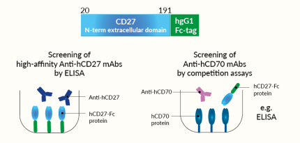
InvivoGen offers hCD27-Fc, a soluble human CD27 chimera protein generated by fusing the N-terminal extracellular domain of human CD27 (aa 20-191) to the N-terminus of a human IgG1 Fc domain with a TEV (Tobacco Etch Virus) sequence linker.
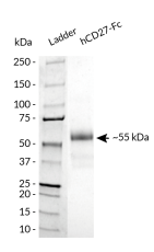
hCD27-Fc has been produced in CHO cells and purified by affinity chromatography. It has an apparent molecular weight of ~55 kDa on an SDS‑PAGE gel.
CD27 background
CD27 is a Type I transmembrane protein and a member of the TNFR family known as the sole receptor for CD70 (aka CD27L). In humans, CD27 is constitutively expressed on the majority of T cells, memory B cells and plasma cells, and natural killer (NK) cells. CD70 is primarily expressed on antigen-presenting cells (APCs). The CD70-CD27 interaction promotes T cell activation and maturation, in concert with the T cell receptor engagement [1]. The CD27/CD70 axis is thus considered a costimulatory immune checkpoint [2].
Applications
- Screening of high-affinity anti-human CD27 monoclonal antibodies by ELISA
- Screening of anti-human CD70 monoclonal antibodies using competition assays
Quality control
- Size and purity confirmed by SDS-PAGE
- Protein validated by ELISA using Anti-hCD27-hIgG1 monoclonal antibody
- Protein validated by flow cytometry using Raji-APC Null cells
References
1. Jacobs, J. et al., 2015. CD70: An emerging target in cancer immunotherapy. Pharmacol Ther 155, 1-10.
2. Flieswasser, T. et al., 2022. The CD70-CD70 axis in oncology: the new kids on the block. J Exp Clin Cancer Res 41, 12.
All InvivoGen products are for internal research use only, and not for human or veterinary use.
InvivoGen also offers:
SPECIFICATIONS
Specifications
NM_001242.4
Sodium phosphate buffer with glycine, saccharose and stabilizing agents
Negative (tested using EndotoxDetect™ assay)
ELISA, flow cytometry
- Screening of high-affinity anti-human CD27 monoclonal antibodies by ELISA
- Screening of anti-human CD70 monoclonal antibodies using competition assays
Each lot is functionally tested and validated.
CONTENTS
Contents
-
Product:hCD27-Fc
-
Cat code:fc-hcd27
-
Quantity:50 µg
1.5 ml of endotoxin-free water
Shipping & Storage
- Shipping method: Room temperature
- -20°C
- Avoid repeated freeze-thaw cycles
Storage:
Caution:
Details
The CD27/CD70 immune checkpoint
The CD27–CD70 axis plays an important role in immune regulation and is considered an immune checkpoint (IC).
The cluster of differentiation CD27 is a member of the TNFR (tumor necrosis factor receptor) superfamily. In humans, CD27 is constitutively expressed on most T cells, memory B cells, plasma cells, and Natural Killer cells [1].
The Cluster of Differentiation CD70 (aka CD27L, TNFSF7) is also a member of the TNFR family and is known as the sole ligand for CD27. Expression of CD70 is tightly controlled and induced transiently in antigen-presenting cells (APCs) following stimuli exposure, such as TFN-α, irradiation, or TLR agonists [2, 3].
The CD27 and CD70 interaction triggers the TRAF2-TRAF6-NF-kB signaling pathway, and in concert with the T cell receptor (TCR) crosslinking, contributes to efficient T cell activation, proliferation, survival, maturation of effector capacity, and T cell memory [2-4].
The CD27/CD70 immune checkpoint in cancer
CD27 is expressed in various types of hematologic cancers. In leukemia, CD27 signaling leads to the induction of different pathways, supporting stemness, tumor cell proliferation, and self-renewal [1]. The efficacy of targeting the CD27/CD70 axis with an agonistic CD27 mAbs was shown in preclinical models of lymphoma, renal cell carcinoma (RCC), breast cancer, and sarcoma [5]. A human mAb directed at CD27 named varlilumab (also CDX-1127, 1F5) has entered clinical trials. It can activate CD27-positive T cells while mediating the killing of CD27-expressing tumor cells [5, 6]. Also, it has successfully completed a phase I/II dose escalation and cohort expansion study (NCT02335918) in combination with nivolumab in different solid malignancies. Further phase I trials with various combinations as well as phase II trials are still ongoing in renal cell carcinoma, squamous cell carcinoma of the head and neck, ovarian and colorectal cancers, and glioblastoma [5].
Constitutive overexpression of CD70 has been described in a range of solid and hematological malignancies, whereby tumor cells have hijacked the CD27-CD70 signaling pathway to facilitate immune evasion in the tumor microenvironment (TME) [7]. They do this by increasing the amount of suppressive regulatory T cells (Tregs), inducing caspase-dependent apoptosis of T cells, and skewing T cells towards exhaustion, to ensure they circumvent the immune response [2]. Additionally, the presence of CD70 on cancer stem cells is a predictive marker for metastasis and poor prognosis [8]. Exploiting CD70-targeting in cancer patients could help eliminate the CD70-expressing cancer cell populations and abrogate the tumor-promoting mechanisms by the CD70-CD27 signaling axis, both in the early stage and advanced disease. Combinatorial approaches with anti-CD70 targeting therapies have proven their potential in both preclinical and clinical settings, using antibody-mediated therapy, chemotherapy, and Chimeric Antigen Receptor (CAR)-T cell therapy [1].
References:
1. Flieswasser, T., Van den Eynde, A., Van Audenaerde, J. et al. 2022. The CD70-CD27 axis in oncology: the new kids on the block. J Exp Clin Cancer Res 41, 12.
2. Jacobs, J. et al. 2015. CD70: An emerging target in cancer immunotherapy. Pharmacol Ther 155, 1-10.
3. Buchan S.L, et al. 2018. The immunobiology of CD27 and OX40 and their potential as targets for cancer immunotherapy. Blood 131(1):39-48.
4. Sanborn RE, et al., 2022. Safety, tolerability and efficacy of agonist anti-CD27 antibody (varlilumab) administered in combination with anti-PD-1 (nivolumab) in advanced solid tumors. J Immunother Cancer. 10(8):e005147.
5. Starzer AM, Berghoff AS., 2020. New emerging targets in cancer immunotherapy: CD27 (TNFRSF7). ESMO Open. 4 (Suppl 3):e000629.
6. Vitale LA, et al., 2012. Development of a human monoclonal antibody for potential therapy of CD27-expressing lymphoma and leukemia. Clin Cancer Res. 18(14):3812-21
7. Jacobs, J. et al. 2018. Unveiling a CD70-positive subset of cancer-associated fibroblasts marked by pro-migratory activity and thriving regulatory T cell accumulation. Oncoimmunology 7, e1440167.
8. Liu, L. et al. 2018. Breast cancer stem cells characterized by CD70 expression preferentially metastasize to the lungs. Breast Cancer 25, 706-716.
DOCUMENTS
Documents
Technical Data Sheet
Validation Data Sheet
Safety Data Sheet
Certificate of analysis
Need a CoA ?


