


LDH-Blue™ Cytotoxicity Assay
| Product | Unit size | Cat. code | Docs. | Qty. | Price | |
|---|---|---|---|---|---|---|
|
LDH-Blue™ Lactate Dehydrogenase (LDH) Activity Assay Kit |
Show product |
200 tests 1000 tests (5 x 200 tests) 10000 tests (50 x 200 tests) |
rep-ldh-1
|
|
Colorimetric LDH release assay for cell death detection

LDH-Blue™ is a high-performance, colorimetric assay for measuring lactate dehydrogenase (LDH) activity in damaged cell supernatant, providing a reliable readout of cell death and a powerful complement to traditional cell viability assays. Designed for simplicity and sensitivity, LDH-Blue™ provides a robust readout of lytic cell death, including necroptosis, pyroptosis, ferroptosis, late apoptosis, and immune cell-mediated cytotoxicity.
Assay principle
LDH is a stable cytoplasmic enzyme released into the culture medium when the plasma membrane is compromised. LDH-Blue™ detects extracellular LDH via a coupled enzymatic reaction that converts a tetrazolium salt into a blue formazan product, proportional to LDH activity. This color change can be quantified using a spectrophotometer at 650 nm.
Key features
- Practical format: Add, Mix, & Read
- Visual results in < 30 min
- Precise and sensitive
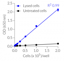
- Absorbance linearity with the number of lysed cells
- No interference with phenol red
- Cost-efficient
All InvivoGen products are for internal research use only, and not for human or veterinary use.
Figures


Specifications
Applications: Lytic cell death monitoring, Compound-mediated cytotoxicity, Cell-mediated cytotoxicity, Antibody-dependent cellular cytotoxicity (ADCC), Pathogen-induced cell death, Genetic screening or gene editing-induced cytotoxicity
Sample types: Cell culture supernatants
Detection method: Colorimetric
Form: Reagent Mix is lyophilized. Assay Buffer, Lysis Buffer, and Stop Solution are liquid.
Quality control: Each lot is functionally tested and validated.
Back to the topContents
LDH-Blue™ is a four-component kit that contains:
– Reagent Mix (lyophilized)
– Assay Buffer
– Lysis Buffer
– Stop Solution
LDH-Blue™ is supplied in three different formats:
- rep-ldh-1: sufficient to monitor LDH activity in 2 x 96-well plates (~200 tests)
- rep-ldh-5: sufficient to monitor LDH activity in 10 x 96-well plates (~1,000 tests)
- rep-ldh-50: sufficient to monitor LDH activity in 100 x 96-well plates (~10,000 tests)
![]()
![]()
![]()
Details
Assay principle

LDH-Blue™ cytotoxicity assay principle
The LDH activity is typically determined by utilizing a coupled enzymatic reaction where LDH oxidizes lactate to pyruvate, converting NAD+ to NADH. Subsequent reduction of tetrazolium salts by NADH and phenazine methosulfate (PMS) leads to the formation of colored formazan. The amount of formazan is directly proportional to cell viability. It can be easily detected with the naked eye and quantified using a spectrophotometer.
InvivoGen's LDH-Blue™ kit contains all the necessary compounds for assessing LDH activity, including Nitro-Blue Tetrazolium (NBT) salts. The NBT reduction produces blue/purple formazan, which can be quantified by measuring the absorbance at 650 nm.
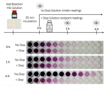
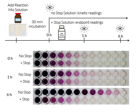
LDH-Blue™ assay at a glance

Back to the top
FAQ
Q: What materials are required to perform the LDH activity reading?
To monitor cytotoxicity using InvivoGen's LDH-Blue™, you will need:
- test and control supernatants
- pipettes and pipette tips, including multi-channel pipette
- reagent reservoir
- flat-bottom transparent 96-well plates
- a spectrophotometer microplate reader capable of reading 650nm absorbance.
Q: What are the storage conditions and stability of LDH-Blue™ reagents?
The kit components are shipped at room temperature. Store the Reagent Mix at -20 °C and protect it from light. Store the Assay Buffer, Lysis Buffer, and Stop Solution at +4 °C. When stored as directed, the kit is stable for up to 1 year from the date of receipt. Once resuspended, the Reagent Mix is stable for 1 week when stored at -20 °C.
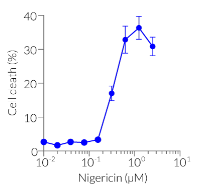
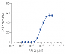
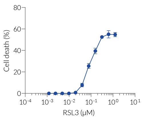
Q: What forms of cell death does LDH-Blue™ detect?
LDH-Blue™ detects lytic cell death associated with plasma membrane disruption, including necroptosis, pyroptosis, ferroptosis, late apoptosis, and immune cell-mediated cytotoxicity. It is not specific to apoptotic pathways without membrane rupture.
Q: Can LDH-Blue™ differentiate between necrosis and apoptosis?
LDH-Blue™ primarily detects cell death associated with membrane integrity loss, such as necrosis or late-stage apoptosis. For early apoptosis detection, complementary assays like Annexin V staining are recommended.
Q: Can I use LDH-Blue™ with immune cell co-cultures?
Absolutely. LDH-Blue™ is ideal for T cell, NK cell, or antibody-mediated cytotoxicity assays. Ensure proper controls for spontaneous and maximum release.
Q: Can LDH-Blue™ be used with low cell number samples, such as 3D microtissues or primary cells?
Yes, LDH-Blue™ is sensitive enough to detect LDH release from samples with low cell numbers, including 3D microtissues and primary cells. Its high sensitivity ensures accurate cytotoxicity assessment in various experimental setups.
Q: Is LDH-Blue™ compatible with high-throughput screening applications?
Absolutely. LDH-Blue™ is designed for use with standard 96-well and 384-well plate formats, making it suitable for high-throughput screening of cytotoxicity in drug discovery and other large-scale studies. You must change the volumes of the supernatant and reagent mix solution (and stop solution) according to the plate format. For a 96-well plate, please see the technical datasheet.
Q: Is the assay compatible with serum-containing media?
Yes, LDH-Blue™ is optimized for use with serum-containing or serum-free media. Background levels may vary slightly with serum type.
Q: Are there any known substances that may interfere with the LDH-Blue™ assay?
Certain substances, such as high concentrations of reducing agents or components that affect enzyme activity, may interfere with the assay. It's advisable to include appropriate controls to account for potential interferences.
Q: Can I store the assay supernatants for later readings?
Depending on your assay, you may want to store the supernatants for later reading. We recommend storing the supernatant at 4°C for up to 48 hours and at -80°C for a longer period. Do not store at -20°C.
Q: What if I observe low absorbance values in my experiment?
There are different possible explanations for low absorbance values:
- The LDH reaction time is too short. If so, do not use the optional Stop Solution and perform kinetic readings of LDH activity.
- The cell culture density is too low. If so, repeat the experiment to optimize cell number.
- The test compound may interfere with the LDH oxydo-reductive reaction (see FAQ above).
- Bubbles are present in the wells when performing the reading. Avoid bubble formation when pipetting and mixing.
Q: What if I observe high variations in replicate values?
One possible reason for the high variation in replicate values could be pipetting errors or insufficient mixing of the Reagent Mix Solution with supernatant samples. We recommend you repeat the experiment using another set of pipettes and/or mix the plate more thoroughly (avoiding bubble formation).
Q: Can I multiplex LDH-Blue™ with other readouts?
Since LDH-Blue™ is non-radioactive and uses absorbance-based detection, it can be multiplexed with fluorescence or luminescence-based assays from the same sample (with appropriate timing). For example, you can use LDH-Blue™ with InvivoGen's Lucia luciferase-based cellular assays: the HMGB1 release reporter cells THP1-HMGB1-Lucia™, the ferroptosis reporter cells HT1080-HMGB1-Lucia™, and the oxidative stress reporter cells HEK-Lucia-Star™ ARE.
Back to the top